A 7-year experience in core needle biopsy of breast lesions: Correlation between imaging and hematoxylin and eosin-stained sections Scientific paper
Article Sidebar
Main Article Content
Abstract
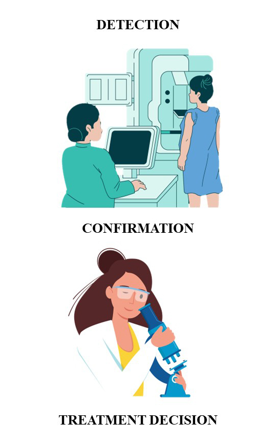
Screening mammography is an imaging procedure which allows breast cancer detection in its early stage. The Breast Imaging and Reporting Data System (BI-RADS) determined six radiological categories for describing lesions. The core needle biopsy (CNB) is minimally invasive procedure that provides pathohistological samples. Via microscopic analysis, samples are categorized into five groups according to the B system for pathohistological report. The aim of the study was to follow the spectrum of pathohistological diagnoses; to define which BI-RADS and core categories are most commonly expressed in certain age groups; and to determine the incidence of histological diagnoses in different BI-RADS categories. The study included 631 patients and data was analysed in order to localise the lesion, BI-RADS and core category and pathohistological diagnosis. Within 631 biopsies, 33 diagnoses were given. In each age group, the findings indicating a high risk for malignancy were the most common (>2 %). The highest percentage of malignant categories was found in patients over the age of 61. Final diagnoses showed a deviation compared to the radiological categories, especially in BI-RADS4 category. Pathohistological diagnosis is always a definite confirmation of a breast lesion type and it has significant contribution to the evaluation of CNB quality.
Downloads
Metrics
Article Details

This work is licensed under a Creative Commons Attribution 4.0 International License.

Authors retain copyright and grant the journal right of first publication with the work simultaneously licensed under a Creative Commons Attribution license 4.0 that allows others to share the work with an acknowledgement of the work's authorship and initial publication in this journal.


References
F. Bray, J. Ferlay, I. Soerjomataram, R. L. Siegel, L. A. Torre, A. Jemal, CA. Cancer. J. Clin. 68 (2018) 394 (https://dx.doi.org/10.3322/caac.21492)
H. G. Welch, C. P. Prorok, A. J. O’Malley, B. S. Kramer, N. Engl. J. Med. 375 (2016) 1438 (https://dx.doi.org/10.1056/NEJMoa1600249)
A.A. Mohamed, Y. Luo, H. Peng, R. C. Jankowitz, S. Wu, J. Digit. Imag. 31 (2018) 387 (https://dx.doi.org/10.1007/s10278-017-0022-2)
H. Pu, J. Peng, F. Xu, N. Liu, F. Wang, X. Huang, Y. Jia, Clin. Breast. Cancer. 20 (2020) 317 (https://dx.doi.org/10.1016/j.clbc.2020.02.009)
N. Houssami, D. Bernardi, F. Caumo, S. Brunelli, C. Fantò, M. Valentini, G. Romanucci, M. A. Gentilini, M. P. Macaskill, Breast 38 (2018) 150 (https://dx.doi.org/10.1016/j.breast.2018.01.002)
R. R. Winkel, M. Euler-Chelpin, E. Lynge, P. Diao, M. Lillholm, M. Kallenberg, J. L. Forman, M. B. Nielsen, W. Y. Uldall, M. Nielsen, I. Vejborg, Cancer. Epidemiol. 49 (2017) 53 (https://dx.doi.org/10.1016/j.canep.2017.05.006)
A. A. Mohamed, W. A. Berg, H. Peng, Y. Luo, R. C. Jankowitz, S. Wu, Med. Phys. 45 (2018) 314 (https://dx.doi.org/10.1002/mp.12683)
M. Wang, X. He, Y. Chang, G. Sun, L. Thabane, Breast 31 (2017) 157 (https://dx.doi.org/10.1016/j.breast.2016.11.009)
M. Von Euler-Chelpin, M. Lillholm, I. Vejborg, M. Nielsen, E. Lynge, Breast. Cancer. Res. 21 (2019) 111 (https://dx.doi.org/10.1186/s13058-019-1203-3)
S. D. Raj, V. Fein-Zachary, P. J. Slanetz, Semin. Ultrasound CT. MR. 39 (2018) 16 (https://dx.doi.org/10.1053/j.sult.2017.08.001)
M. Sirous, S. P. Shahnani, A. Sirous, Adv. Biomed. Res. 7 (2018) 56 (https://dx.doi.org/10.4103%2Fabr.abr_161_17)
A. Gastounioti, E. F. Conant, D. Kontos, Breast. Cancer. Res. 18 (2016) 91 (https://dx.doi.org/10.1186/s13058-016-0755-8)
E. Łukasiewicz, A. Ziemiecka, W. Jakubowski, J. Vojinovic, M. Bogucevska. J. Ultrason. 17 (2017) 267 (https://dx.doi.org/10.15557/jou.2017.0039)
I. Chakrabarti, J. Cytol. 35 (2018) 176 (https://dx.doi.org/10.4103%2FJOC.JOC_35_18)
C. E. Edmonds, L. R. Lamb, S. F. Mercaldo, D. A. Sippo, K. S. Burk, C. D. Lehman, Am. J. Roentgenol. 214 (2020) 240 (https://dx.doi.org/10.2214/AJR.19.21778)
S. Kulkarni, S. Murchite, A. Patil, J. Surg. Res. 5 (2022) 221 (https://dx.doi.org/10.26502/jsr.10020215)
J. C. Litherland, Clin. Radiol. 57 (2002) 81 (https://dx.doi.org/10.1053/crad.2001.0875)
C. A. Pistolese, D. Tosti, D. Citraro, F. Ricci, C. Di Stefano, F. Lamacchia, D. Ferrari, R. Floris, Ultrason Med Biol. 45 (2019) 78 (https://dx.doi.org/10.1016/j.ultrasmedbio.2018.09.004)
W. A. Berg, J. Breast Imag. 3 (2021) 527 (https://dx.doi.org/10.1093/jbi/wbab060)
A. Hamidinekoo, E. Denton, A. Rampun, K. Honnor, R. Zwiggelaar, Med. Imag. Anal. 47 (2018) 45 (https://dx.doi.org/10.1016/j.media.2018.03.006)
G. Aresta, T. Araujo, S. Kwok, S. S. Chennamsetty, M. Safwan, V. Alex, Med. Imag. Anal. 56 (2019) 122 (https://dx.doi.org/10.1016/j.media.2019.05.010).